
 Product sheet · Quantel Medical Compact Touch
Compact Touch®
Tablet-sized A/B/P ultrasound platform: B-scan imaging, biometry with IOL calculation, and pachymetry.
Compact Touch is a touchscreen ophthalmic ultrasound platform weighing under 4 kg. It combines a new-generation 15 MHz B-scan probe, ultrasound biometry with IOL calculation for all eye types, and optional pachymetry, with DICOM and EMR connectivity.
- < 4 kgtotal weight
- +30%B-scan resolution
- 12IOL calculation formulas
Key features
- 01
Sharper B-scan
New-generation 15 MHz B probe with 30% higher resolution for better visualization of the eye and orbit. The small probe is easy to handle.
- 02
Biometry in B-mode (exclusive)
Measures axial length exactly at the macula, even in unusual eyes such as long myopic eyes with staphylomas, while ruling out other posterior pole pathology.
- 03
Biometry for every eye
Ultrasound biometry works regardless of cataract density or media opacity, with precision similar to optical biometry (0.03 mm in immersion). 12 IOL formulas are included, with post-refractive options.
- 04
ProBeam™ fixation (option)
An exclusive biometry probe that projects a laser fixation point for the other eye, making measurement easier and more precise.
- 05
Pachymetry with IOP correction (option)
Precision of ±5 µm over a 200–999 µm range, with Ehlers, Doughty and Dresdner IOP correction tables.
- 06
Truly compact
3.5 kg, fanless and silent, with a folding carry handle. It mounts by VESA on a wall, arm or cart (option).
Who it's for
Ideal for
Clinical uses
- Posterior segment and orbit B-scan imaging
- Axial length biometry and IOL calculation
- Biometry in dense cataract and opaque media
- Corneal pachymetry for refractive surgery and glaucoma
Technical specifications
B mode
- Grey levels
- 256
- Adjustable gain
- 20 to 110 dB
- Time Gain Control (TGC)
- 0 to 30 dB
- Dynamic range adjustment
- 25 to 90 dB
- Storage
- Still images and video sequences (up to 40 seconds) on hard disk
- Image post-processing tools
- Calipers, areas, markers, comment
15 MHz B probe
- Transducer frequency
- 15 MHz
- Angle of exploration
- 50°
- Depth of exploration
- 60 mm (2.36″)
- Focus
- 24 mm (0.94″)
- Axial resolution
- 115 µm
- Lateral resolution
- 400 µm
- Frame rate acquisition
- Up to 16 Hz
Biometry (11 MHz probe)
- Adjustable gain
- 20 to 110 dB
- Time Gain Control (TGC)
- 0 to 30 dB
- Transducer frequency
- 11 MHz
- Tip diameter
- 7 mm (0.28″)
- Electronic resolution
- 0.03 mm (0.002″)
- Depth
- 60 mm (2.4″) on 1536 points
- Techniques
- Contact and immersion compatible
- Aiming beam
- LED or ProBeam™ laser pointer (option)
- Axial length measurement
- Ultrasound velocity adjustable per segment (anterior chamber, lens, vitreous) and for IOL and vitreous material
Built-in pattern recognition: phakic, aphakic, PMMA, acrylic and silicone pseudo-phakic eyes
Automatic standard deviation and average total length (series of 10 measurements)
Acquisition modes: automatic, auto + save, manual
Automatic detection of scleral spike - IOL calculation
- SRK-T, SRK-II, Holladay, Binkhorst-II, Hoffer-Q, Haigis
- Post-refractive calculation
- Pre-op and post-op refraction and keratometry
6 methods for keratometric correction and implant calculation: history derived, refraction derived, contact lens method, Rosa regression, Shammas regression, Double K/SRK-T (Dr. Aramberri's formula) - IOL display
- 9 values bracketed for desired ametropia for each IOL (0.25 D or 0.50 D steps); simultaneous display of 4 IOL calculations
Pachymetry (option)
- Transducer frequency
- 20 MHz
- Tip diameter
- 1.2 mm (0.05″)
- Method
- Contact
- Convergence
- 0.5 mm (0.02″) from the tip
- Angle
- 45°
- Measurement range
- 200 to 999 µm
- Number of measurements
- 1 to 10
- Precision
- ± 5 µm
- Velocity
- Adjustable
- Methods
- Central measurement or cartographic map (automatic, continuous, scanning)
- Cartographic maps
- User, 9C8L, 9C4L, 5C8L, 5C4L, 9C, 5C, 8L, 4L
- IOP correlation tables
- Ehlers, Doughty, Dresdner and unlimited user-defined tables
- Bias correction
- Up to 120%
Data management
- Database
- Built-in physician and patient database
- Export
- Still images and video sequences
- Reports
- Customisable digital and printed report
- Connectivity
- DICOM compatible (Worklist, Storage, Print) (option); cineloops in DICOM format; EMR compatible
- Printing
- Compatible with PC and USB video printers
General
- Display
- Back-lit LCD colour touch screen, 1024 × 768 px
- Touch screen dimensions
- 21 cm (W) × 16 cm (H) / 8.3″ × 6.3″
- Overall dimensions
- 26.8 cm (W) × 4.0 cm (D) × 24.6 cm (H)
10.6″ (W) × 1.6″ (D) × 9.7″ (H) - Weight
- 3.5 kg (7.7 lbs)
- Ports
- 4 USB, 1 Ethernet, 1 HDMI
- Power supply
- 100–240 Vac ±10% single phase with earth, 50/60 Hz, 60 W max
- Included
- Footswitch, Bluetooth mouse
- Options
- External PC printer (USB or Wi-Fi), USB video printer, cart, articulating arm (wall or table)
Source: Quantel Medical brochure. Specifications are subject to change without notice. Open the original brochure.
Images
Related products
-
 Quantel Medical
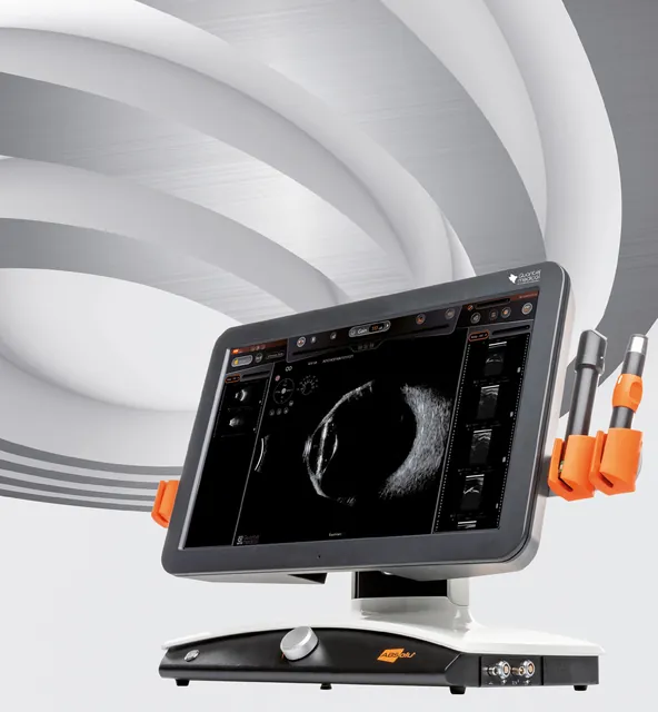
ABSolu®
Premium A/B/S/UBM ultrasound platform: whole-eye imaging, UBM for glaucoma, biometry and standardized echography.
-
 OCULUS
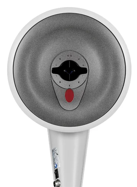
Pentacam® Family
The gold standard in anterior segment tomography: Scheimpflug imaging, optical biometry, wavefront and cornea OCT in five models.
-
 Heidelberg Engineering
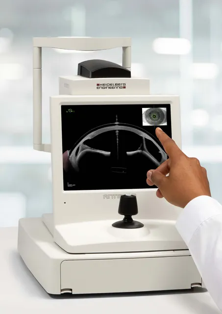
ANTERION®
All-in-one swept-source OCT for the anterior segment: biometry and IOL calculation, corneal tomography, AC metrics and imaging.