
 Product sheet · Quantel Medical ABSolu
ABSolu®
Premium A/B/S/UBM ultrasound platform: whole-eye imaging, UBM for glaucoma, biometry and standardized echography.
ABSolu is Quantel Medical's top-end ophthalmic ultrasound platform. A 5-ring annular 20 MHz probe images the whole eye in exceptional detail, a 50 MHz UBM probe shows structures behind the iris for glaucoma and ICL sizing, and A-scan, B-mode biometry and standardized A-mode complete the platform. It runs on a 21″ Full HD DICOM-calibrated screen.
- +70%depth of field (annular probe)
- 35 µmUBM axial resolution
- 21″Full HD DICOM display
Key features
- 01
Annular 20 MHz probe
Five concentric transducers in one probe give +70% depth of field and 25% better lateral resolution (250 → 200 µm), showing vitreous, retina and beyond in one scan.
- 02
Optimized UBM
50 MHz linear-scan UBM shows structures behind the iris that other technologies cannot see. Clearscan™ compatible for fast, comfortable exams.
- 03
Glaucoma module
Semi-automatic angle quantification (AOD, TIA, IT, ARA, TISA, LV) to understand iris, lens and ciliary body mechanisms in glaucoma patients.
- 04
STS module and ICL sizing (option)
Automatic sulcus-to-sulcus, lens curvature and anterior chamber depth in a single scan. With GURU Export it sends images to the ICLguru platform for ICL sizing.
- 05
Patented motion sensor
The B15, B20-5A and UBM probes show the probe position on the eye and the explored area in real time (IMUv®).
- 06
Standardized echography (option)
The only ultrasound platform meeting Prof. Ossoinig's criteria. S-mode supports diagnosis of tumours, retinal/vitreous membrane detachment and Graves' disease.
- 07
DICOM-grade display
A world first in ophthalmic ultrasound: a Full HD greyscale screen compliant with DICOM part 14, for consistent, reliable image interpretation.
Who it's for
Ideal for
Clinical uses
- Posterior segment and orbit imaging
- UBM of the anterior segment and angle
- Glaucoma mechanism assessment (angle closure, pupillary block)
- Sulcus-to-sulcus measurement and ICL sizing
- Axial length biometry and IOL calculation
- Tumour diagnosis and Graves' disease (standardized A-mode)
Technical specifications
B-scan modes
- Grey levels
- 256
- Adjustable gain
- 20 to 110 dB
- Adjustable Time Gain Control (TGC)
- 0 to 30 dB
- Adjustable dynamic range
- 25 to 90 dB (15 and 50 MHz); 80 dB for 20 MHz 5A
- Image post-processing tools
- Filters (algorithm and colours), calipers, areas, angles, markers, comments
- Glaucoma quantifying semi-automated tools
- AOD 500 & 750, TIA, IT 750 & 2000, ARA 500 & 750, TISA 500 & 750, LV
- Cineloop in B mode
- Up to 400 images
Posterior pole: magnetic 15 MHz probe
- Transducer frequency
- 15 MHz
- Angle of exploration
- 50°
- Depth of exploration
- 60 mm (2.36″)
- Focus
- 24 mm (0.94″)
- Depth of field
- 12 mm (0.47″)
- Axial resolution
- 115 µm
- Lateral resolution
- 400 µm
- Frame rate acquisition
- Up to 16 Hz
- Probe localization
- Accelerometer – IMUv®
Posterior pole: magnetic annular 5-ring 20 MHz probe
- Transducer frequency
- 20 MHz – annular 5 rings
- Angle of exploration
- 50°
- Depth of exploration
- 40 mm (1.57″)
- Focus
- 22 mm (0.87″)
- Depth of field
- 20 mm (0.79″)
- Axial resolution
- 80 µm
- Lateral resolution
- 200 µm
- Frame rate acquisition
- Up to 16 Hz
- Probe localization
- Accelerometer – IMUv®
UBM & anterior segment: magnetic 50 MHz UBM probe
- Transducer frequency
- 50 MHz, linear scanning
- Linear transducer movement
- 16 mm (0.63″)
- Focus
- 10 mm (0.39″)
- Axial resolution
- 35 µm
- Lateral resolution
- 60 µm
- Probe localization
- Accelerometer – IMUv®
- Options
- STS module, GURU Export
Standardized A mode (option)
- Amplifier
- Digitally programmed S-shaped amplifier characteristics and comprehensive design criteria for standardized echography and tissue differentiation according to Prof. Dr. Karl C. Ossoinig
- Tissue sensitivity
- Automatic tissue sensitivity determination with specific gain value recorded
- Diagnosis functions
- LESION Q-I, Retina A1, Retina QII, muscle profile with optic nerve measurements
- Probe frequency
- 8 MHz parallel beam
- Cineloop in A mode
- Up to 400 images
- Depth
- Orbit 80 µs, eye 40 µs, zoom 20 µs
- Measurement
- Distance measurement between 2 gates with adjustable velocity
Biometry (11 MHz probe)
- Adjustable gain
- 20 to 110 dB
- Adjustable TGC
- 0 to 30 dB
- Transducer frequency
- 11 MHz
- Tip diameter
- 7 mm (0.28″)
- Electronic resolution
- 0.04 mm (0.0016″)
- Depth of exploration
- 40/80 mm (1.57″/3.15″) on 2048 points
- Aiming beam
- LED or ProBeam™ laser beam
- Techniques
- Contact and immersion compatible
- Axial length measurement
- Ultrasound velocity adjustable per segment (anterior chamber, lens, vitreous) and for IOL and vitreous material
Built-in pattern recognition: phakic, dense/long, aphakic, PMMA, acrylic and silicone pseudo-phakic eyes
Acquisition modes: automatic, auto + save, manual
Automatic detection of scleral spike
Automatic standard deviation and average total length (series of 10 measurements) - IOL calculation
- SRK-T, SRK 2, Holladay, Binkhorst-II, Hoffer-Q, Haigis
- Post-refractive calculation
- Pre-op and post-op refraction and keratometry
6 methods for keratometric correction and implant calculation: history derived, refraction derived, contact lens method, Rosa regression, Shammas regression, Double K/SRK-T (Dr. Aramberri's formula) - IOL display
- 9 values bracketed for desired ametropia for each IOL (0.25 D or 0.50 D steps); simultaneous display of 4 IOL calculations
Data management
- Database
- Built-in physician and patient database; no restriction on number of exams per patient
- Export
- Still images and video sequences
- Reports
- Customizable digital and printed reports
- Connectivity
- DICOM (option) and/or EMR compatible
- Printing
- Compatible with PC, USB video and DICOM printers
General
- Screen
- 21″ HD (1920 × 1080), greyscale display compliant with DICOM part 14
- Languages
- Chinese, English, French, German, Japanese, Polish, Spanish
- Connections
- 5 USB ports (1 on the base, 4 under the screen), HDMI and Ethernet
- Computer
- Windows 11 LTSC; HDD 2 TB, SSD 128 GB, RAM 16 GB
- Power supply
- 80–264 Vac, 47/63 Hz, 65 VA max
- Overall dimensions
- Height 445 mm (17.52″), depth 285 mm (11.22″), width 545 mm (21.46″) without probe holders, 840 mm (33.07″) with all probes
- Weight
- 11.5 kg (25.35 lbs) without probes
Source: Quantel Medical brochure. Specifications are subject to change without notice. Open the original brochure.
Images
Related products
-
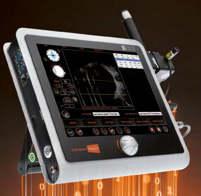 Quantel Medical
Compact Touch®
Tablet-sized A/B/P ultrasound platform: B-scan imaging, biometry with IOL calculation, and pachymetry.
-
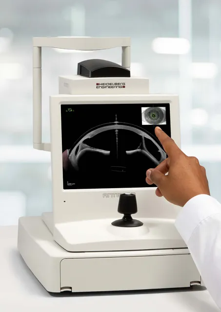 Heidelberg Engineering
ANTERION®
All-in-one swept-source OCT for the anterior segment: biometry and IOL calculation, corneal tomography, AC metrics and imaging.
-
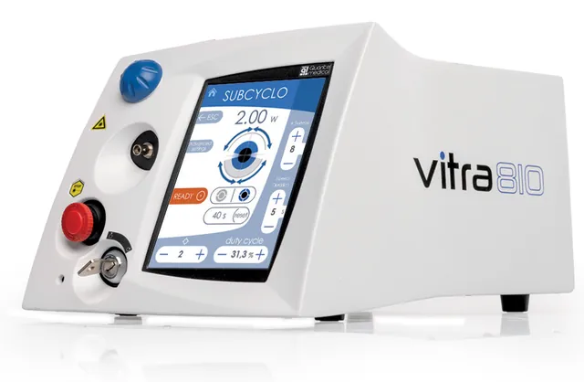 Quantel Medical
VITRA 810 for SubCyclo®
810 nm diode laser for SubCyclo® non-destructive glaucoma treatment, plus standard 810 nm retina treatments.