
 Product sheet · Heidelberg Engineering ANTERION
ANTERION®
All-in-one swept-source OCT for the anterior segment: biometry and IOL calculation, corneal tomography, AC metrics and imaging.
ANTERION is an upgradeable all-in-one platform for the anterior segment, built on long-wavelength (1300 nm) swept-source OCT. One small-footprint device covers biometry and IOL power calculation, corneal topography and tomography, anterior chamber metrics and high-resolution imaging. Because the OCT images behind every measurement are shown, clinicians can see what they measure.
- 1300 nmswept-source OCT
- ~16,000corneal data points
- < 1 sfor 65 corneal B-scans
Key features
- 01
One device, whole anterior segment
Biometry including axial length, cornea exam, anterior segment imaging and patient consultation on one platform. It saves time and space and is easy to delegate.
- 02
Cataract planning with fewer surprises
True measurements of all refractive surfaces and the full corneal power (~16,000 data points). Spheric and toric IOL calculation with Barrett Suite, Haigis, Holladay 1, SRK/T, Hoffer® Q and OKULIX ray tracing.
- 03
Comprehensive cornea analysis
65 OCT B-scans in under one second. OU View differential maps show asymmetry, and Follow-Up View tracks surgical outcomes and disease.
- 04
Ectasia screening with SCORE
The SCORE Analyzer (by Dr. Gatinel and Dr. Saad) plus Ectasia View and the Epithelial Thickness Module support keratoconus screening and refractive surgery planning.
- 05
Angle assessment in 360°
Automatic scleral spur detection and 360° angle graphs complement gonioscopy for angle-closure assessment, along with AC volume, lens vault and ICL vault.
- 06
Expand as you grow
Start with the Imaging App and add the Cataract, Cornea and Metrics Apps when needed.
Who it's for
Ideal for
Clinical uses
- Biometry and IOL power calculation (including toric and post-refractive)
- Keratoconus and ectasia screening
- Refractive surgery planning (epithelial thickness)
- Angle-closure assessment
- Phakic IOL / ICL planning and vault measurement
- Pre- and post-operative comparison (cataract, DMEK, glaucoma surgery)
ANTERION apps
Imaging App
Anterior chamber and angle imaging, corneal and scleral imaging, visualization of the lens and both surfaces, customizable scan patterns, peripheral imaging.
Cataract App (optional)
Axial length, lens thickness, aqueous depth, central corneal thickness, total corneal power, total corneal wavefront, spheric and toric IOL calculator, IOL power prediction, IOL database import, OKULIX ray tracing.
Cornea App (optional)
Corneal topography and tomography, pachymetry, total corneal power, anterior axial curvature, corneal wavefront, differential maps, progression analysis, Ectasia View with SCORE analyzer, Epithelial Thickness Module.
Metrics App (optional)
Anterior chamber angle assessment, 360° graphs of angle parameters, anterior chamber volume, lens vault, lens thickness, free-hand measurements, automatic scleral spur detection.
Technical specifications
Biometry measurement ranges
- Central corneal thickness
- 300 – 1700 µm
- Anterior chamber depth
- 1.50 – 4.80 mm
- Lens thickness
- 2.40 – 6.50 mm
- Axial length
- 14.00 – 32.00 mm
- SimK mean anterior (3 mm)
- 6.00 – 110.00 D
- K mean posterior (3 mm)
- −14.80 – −0.70 D
- Astigmatism anterior
- 0.00 – 15.50 D
- Astigmatism posterior
- −2.10 – 0.00 D
- White-to-white
- 9.40 – 15.30 mm
- Pupil diameter
- 0.20 – 14.10 mm
Technology and software
- Technology
- Swept-source OCT, long wavelength (1300 nm)
- Corneal acquisition
- 65 OCT B-scans in less than one second, more than 16,000 data points of the anterior and posterior corneal surface
- IOL formulas
- Barrett Suite (Universal II, Toric, True-K, True-K Toric), Haigis, Holladay 1, SRK/T, Hoffer® Q, OKULIX ray tracing
- IOL calculation
- Spheric and toric; import options for IOL databases
- Ectasia analysis
- Ectasia View with SCORE (Screening Corneal Objective Risk of Ectasia) Analyzer
- Epithelium
- Epithelial Thickness Module: epithelial and stromal thickness maps and parameters
- Angle analysis
- Automatic scleral spur detection; six OCT images or 360° graph of angle parameters
- Views
- OU View (left/right comparison with differential maps), Follow-Up View, trend analysis
- Modularity
- Imaging App + optional Cataract App, Cornea App and Metrics App
Source: Heidelberg Engineering brochure. Specifications are subject to change without notice. Open the original brochure.
Images
Related products
-
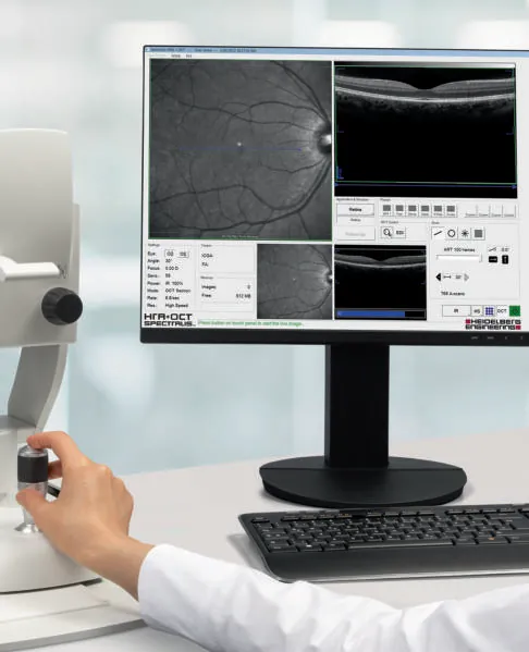 Heidelberg Engineering
SPECTRALIS®
Multimodal imaging platform for the posterior segment: SD-OCT, cSLO, angiography and more, with TruTrack eye tracking.
-
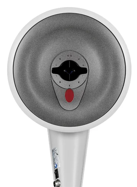 OCULUS
Pentacam® Family
The gold standard in anterior segment tomography: Scheimpflug imaging, optical biometry, wavefront and cornea OCT in five models.
-
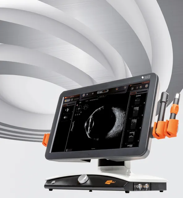 Quantel Medical
ABSolu®
Premium A/B/S/UBM ultrasound platform: whole-eye imaging, UBM for glaucoma, biometry and standardized echography.