
 Product sheet · Heidelberg Engineering SPECTRALIS
SPECTRALIS®
Multimodal imaging platform for the posterior segment: SD-OCT, cSLO, angiography and more, with TruTrack eye tracking.
SPECTRALIS combines confocal scanning laser ophthalmoscopy (cSLO), high-resolution spectral-domain OCT and patented real-time image processing for outstanding image quality and reproducibility. It is modular and upgradeable: start with OCT, then add autofluorescence, MultiColor, FA/ICGA, OCT angiography, widefield, glaucoma and anterior segment modules as the practice grows, keeping all patient data for follow-up.
- 125 kHztop OCT scan speed
- 100B-scans averaged live
- 5.7 µmOCTA lateral resolution
- 55°widefield OCT and fundus
Key features
- 01
TruTrack Active Eye Tracking
A second laser actively tracks the fundus during OCT scanning, freezing the retina even if the patient blinks or moves. Often imitated, never duplicated.
- 02
Reliable follow-up
AutoRescan places follow-up scans in exactly the same anatomic location, so real change can be told apart from noise. Up to 100 B-scans are averaged live.
- 03
SHIFT: three scan speeds
125 kHz for up to 30% faster OCT angiography, 85 kHz for balanced everyday imaging, and 20 kHz for high sensitivity in eyes with media opacities.
- 04
True multimodal imaging
Infrared, blue reflectance, BluePeak autofluorescence, MultiColor, FA, ICGA and OCTA (5.7 µm lateral resolution) on one device, with no moving the patient between rooms.
- 05
Glaucoma Module Premium Edition
Personalized analysis of the optic nerve head, RNFL and macular ganglion cells using the Anatomic Positioning System, with the Hood Glaucoma Report at a glance.
- 06
Future-proof investment
Many modules can be added later without buying a new device, preserving patient data. The Flex Module images patients lying down or unable to use a headrest.
Who it's for
Ideal for
Clinical uses
- Diagnosis and monitoring of AMD, diabetic macular edema and other retinal disease
- Glaucoma diagnosis and progression (ONH, RNFL, ganglion cells)
- Fluorescein and ICG angiography
- OCT angiography of retinal capillaries
- Geographic atrophy (BluePeak autofluorescence)
- Anterior segment OCT (cornea, sclera, angle)
- Neuro-ophthalmic axonal analysis (Nsite Analytics)
SPECTRALIS configurations
SPECTRALIS OCT
Compact version with a small headrest and joystick trigger for fast capture and a reduced footprint. Easy for less experienced users.
SPECTRALIS HRA+OCT
The plus version, with angiography (FA standard), BluePeak and the pan-and-tilt camera head for challenging patients and peripheral imaging.
SPECTRALIS HRA
Angiography and fundus imaging platform, upgradeable to HRA+OCT.
Technical specifications
Configuration options
| Feature | SPECTRALIS OCT | SPECTRALIS HRA+OCT | SPECTRALIS HRA |
|---|---|---|---|
| Fundus: Infrared Reflectance | Standard | Standard | Standard |
| Fundus: BluePeak (autofluorescence) | Optional | Standard | Standard |
| Fundus: MultiColor | Optional | Optional | Optional |
| Fundus: High Magnification Module | Optional | Optional | Optional |
| OCT: Posterior Segment | Standard | Standard | After upgrade to HRA+OCT |
| OCT: Anterior Segment | Optional | Optional | After upgrade to HRA+OCT |
| OCT: Glaucoma Module Premium Edition | Optional | Optional | After upgrade to HRA+OCT |
| OCT: Nsite Analytics | Optional | Optional | After upgrade to HRA+OCT |
| OCT scan speed: 20 kHz (high sensitivity) | Optional | Optional | After upgrade to HRA+OCT |
| OCT scan speed: 85 kHz | Optional | Optional | After upgrade to HRA+OCT |
| OCT scan speed: 125 kHz | Optional | Optional | After upgrade to HRA+OCT |
| Angiography: Fluorescein Angiography | After upgrade to HRA+OCT | Standard | Standard |
| Angiography: ICG Angiography | After upgrade to HRA+OCT | Optional | Optional |
| Angiography: OCT Angiography | Optional | Optional | After upgrade to HRA+OCT |
| Widefield Imaging | Optional | Optional | Optional |
| Ultra-Widefield Imaging | After upgrade to HRA+OCT | Optional | Optional |
| Hardware: Flex Module | Optional | Optional | Optional |
| Hardware: Panning Camera | Optional | Standard | Standard |
Some options can be added anytime; others are only available at initial equipment purchase.
Core technologies and modules
- Imaging technology
- Confocal scanning laser ophthalmoscopy (cSLO) + spectral-domain OCT (SD-OCT), with real-time image processing
- Eye tracking
- TruTrack Active Eye Tracking (patented), using a second laser beam to track the fundus during OCT scanning
- Image averaging
- Up to 100 B-scans averaged live, including in volume scans
- Follow-up
- AutoRescan: follow-up scans in the same anatomic location as previous exams
- Segmentation
- Visualization of 15 structures and segmentation of retinal layers
- OCT scan speeds (SHIFT)
- 20 kHz (high sensitivity), 85 kHz (balanced), 125 kHz (up to 30% faster OCTA acquisition vs 85 kHz)
- Enhanced depth modes
- EDI and EVI modes for the outer retinal layers and the vitreous
- OCT Angiography
- Lateral resolution of 5.7 µm; 3D representation of retinal vascular flow
- Widefield Imaging Module
- 55° field of view for OCT and all simultaneous fundus imaging modalities
- Ultra-Widefield Imaging Module
- Evenly illuminated, undistorted cSLO angiography from the macula through the periphery in one acquisition
- Scanning laser angiography
- FA and ICGA images and videos, acquired simultaneously and in combination with OCT
- Anterior Segment Module
- High-resolution OCT of the cornea, sclera and anterior chamber angles, angle to angle in one shot
- Glaucoma Module Premium Edition
- ONH, RNFL and macular ganglion cell layer analysis; Anatomic Positioning System; Hood Glaucoma Report
- Nsite Analytics
- Neuronal and axonal analysis within the retina, with colour-scheme classification and change over time
- High Magnification Module
- Non-invasive high-resolution imaging of retinal microstructures
- Flex Module
- SPECTRALIS imaging for patients in a supine position or unable to use a traditional headrest
Source: Heidelberg Engineering brochure. Specifications are subject to change without notice. Open the original brochure.
Images
Related products
-
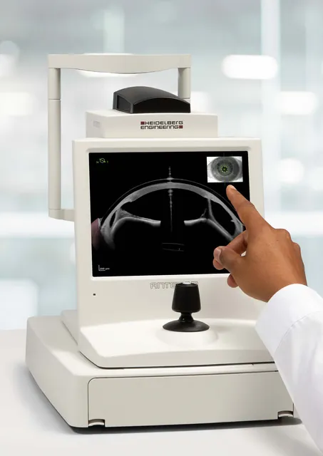 Heidelberg Engineering
ANTERION®
All-in-one swept-source OCT for the anterior segment: biometry and IOL calculation, corneal tomography, AC metrics and imaging.
-
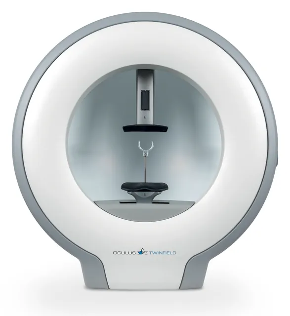 OCULUS
Twinfield® 3
Full-field bowl perimeter for static and kinetic visual field testing, with glaucoma analysis built in.
-
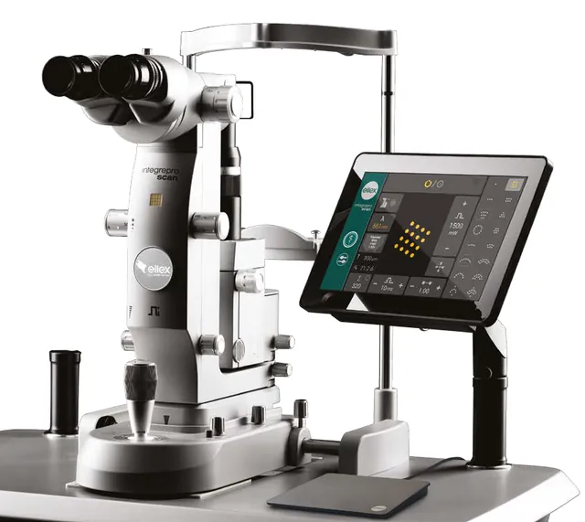 Ellex
Integre Pro & Integre Pro Scan
All-in-one retinal photocoagulation laser and slit lamp, with single-spot and pattern scanning modes.