
 Product sheet · Heidelberg Engineering OPERA
Heidelberg OPERA
Fully digital surgical microscope platform for ophthalmic surgery: see clearly, work naturally, teach together.
Heidelberg OPERA is a fully digital ophthalmic surgical microscope platform. Unlike conventional optical or hybrid systems it is digital by design: two 4K-UHD cameras, 3D 4K monitors and freely positionable digital binoculars give enhanced visualization, an upright working posture, and a shared 2D/3D view with live annotations for the whole OR team. Its modular architecture is built to take on future technologies as digital surgery advances.
- 2× 4KUHD cameras
- 6×motorized optical zoom
- 200 mmworking distance
- Up to 4wireless observer displays
Key features
- 01
Digital by design
Not an optical or hybrid microscope with a camera added: a fully digital platform for enhanced visualization, better ergonomics, teaching and collaboration in the OR.
- 02
Premium digital image quality
Two 4K-UHD cameras with excellent colour, contrast and resolution, enhanced depth of field and advanced digital image enhancement. Digital brightness allows lower illumination, supporting patient comfort.
- 03
Ergonomics for long surgical careers
3D monitors for straight-ahead viewing, freely positionable digital binoculars and a C-shaped suspension arm support an upright posture and reduce neck and back strain.
- 04
Designed for teaching
The whole OR team shares the same real-time 2D/3D view. Assist Mode annotations, 4K-UHD recording and wireless streaming to up to 4 observer displays extend learning beyond the OR.
- 05
Intuitive control
Control by touchscreen or footswitch, with full integration of OCULUS BIOM® fundus viewing systems, including footswitch control.
- 06
Future-ready platform
A modular architecture designed to integrate new technologies, such as swept-source intraoperative OCT (1060 nm, 70,000 A-scans per second). The iOCT module is in development and not yet commercially available.
Who it's for
Ideal for
Clinical uses
- Anterior segment surgery, including cataract surgery and ICL implantation
- Posterior segment (vitreoretinal) surgery
- Surgical teaching with live annotations
- Recording, review and streaming of procedures
Viewing options
Digital binoculars
Freely positionable digital binoculars for surgeons who prefer an eyepiece view, while keeping an upright posture.
55″ 3D 4K monitor
Large heads-up display for straight-ahead 3D viewing, shared with the whole OR team.
32″ 3D 4K monitor
Heads-up display mounted to the microscope stand and positioned in front of the surgeon on the C-arm.
Technical specifications
Display and imaging
- Display options for surgeons
- Digital binoculars
55″ 3D 4K monitor
32″ 3D 4K monitor (mounted to the microscope stand) - Display options for observers
- Up to 4 via wireless transmitter (2D/3D, up to 4K-UHD resolution)
1× 3D via HD-SDI or 2× 2D via HD-SDI - Camera sensor resolution
- 2× 4K-UHD cameras (3840 × 2160 pixels)
- Recording
- 4K-UHD (3840 × 2160), 60 fps
Full-HD (1920 × 1080), 60 fps
Full-HD (1920 × 1080), 30 fps
2D & 3D - Surgical annotations
- Yes
- Video editing
- Yes
Optics and illumination
- Magnification
- Motorized optical 6× zoom
50% digital zoom - Focus range
- 90 mm (in 0° position)
- Working distance
- 200 mm
- Aperture
- Motorized, 9 positions
- Red reflex illumination
- LED
- Surrounding field illumination
- LED
- Built-in keratoscope
- LEDs
- Digital filters
- Color filters, contrast enhancement, sharpness, digital brightness
- Physical filters
- Retina protection filter (built-in)
Retina protection plate (built-in)
532 nm laser filter (built-in)
Connectivity and compatibility
- Interfaces
- 1× HDMI optical output
2× HD-SDI in
2× HD-SDI out
Wireless video transmitter
USB 3.2 gen 2 - Compatible fundus viewing systems
- OCULUS BIOM® 6
OCULUS BIOM® 5ml/cl
OCULUS BIOM® ready/ready+
Full microscope integration, including footswitch control
Dimensions
- Microscope stand height
- 2250 mm
- Microscope stand base
- 750 × 750 mm
- Maximum arm reach
- 1700 mm
- Maximum optics height
- 1650 mm
- Movement
- Arm rotation ± 270°
Optics tilt −45° / +90°
Stand rotation without stop - Monitor cart
- Monitor width 1262 mm, height 1737 mm
Base 621 × 718 mm
Manufacturer
- Legal manufacturer
- Munich Surgical Imaging GmbH, Munich, Germany
- Distributed by
- Heidelberg Engineering GmbH, Heidelberg, Germany
Source: Heidelberg Engineering brochure. Specifications are subject to change without notice. Open the original brochure.
Images
Related products
-
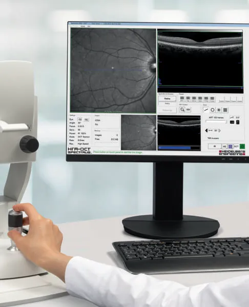 Heidelberg Engineering
SPECTRALIS®
Multimodal imaging platform for the posterior segment: SD-OCT, cSLO, angiography and more, with TruTrack eye tracking.
-
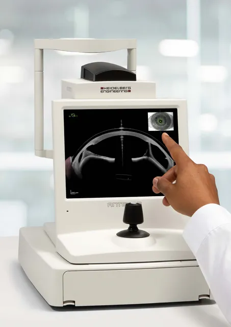 Heidelberg Engineering
ANTERION®
All-in-one swept-source OCT for the anterior segment: biometry and IOL calculation, corneal tomography, AC metrics and imaging.
-
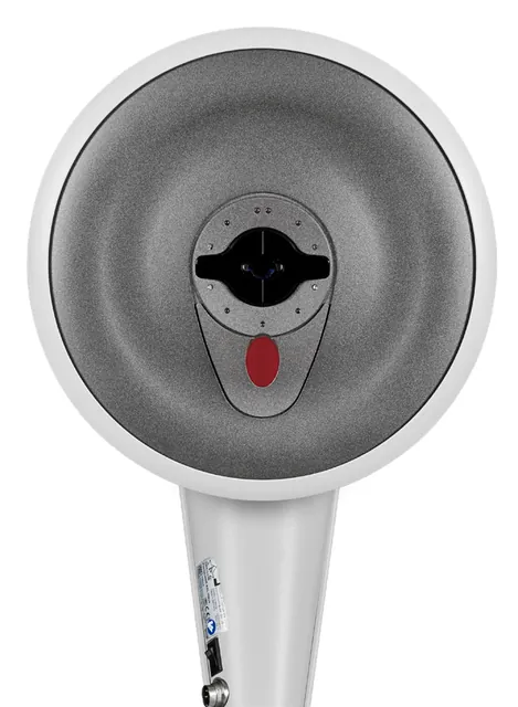 OCULUS
Pentacam® Family
The gold standard in anterior segment tomography: Scheimpflug imaging, optical biometry, wavefront and cornea OCT in five models.